In our lab, we use neurospheres, round structures made of thousands of living neurons, which we train for computations.
How do we know they are neurons?
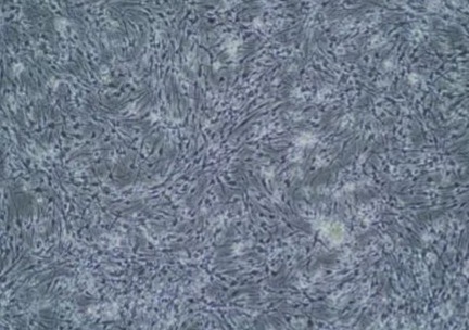
In the lab, all cells look similar. If you look at them with the naked eye, usually you can see some ‘dirt’ on the laboratory glasses, nothing more, as they are too small.
If you look under the microscope, you can see some structure.
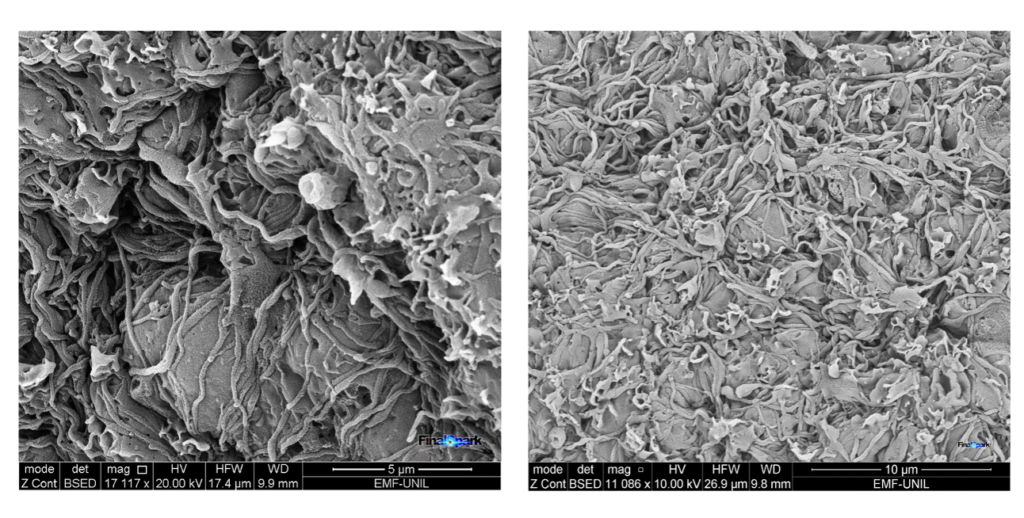
You can go even deeper in your analysis and increase the resolution of a microscope to get very fine detail. Here are the pictures of our neurospheres taken under an electron microscope. We can see quite a lot of details of their shape (morphology).
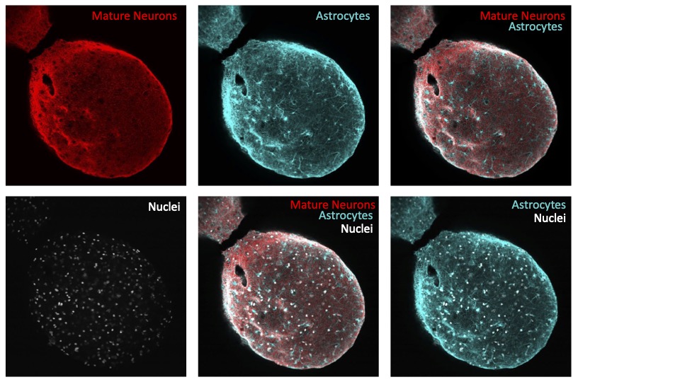
To make sure that we have real neurons in our lab, we can detect some specific molecules which occur only in neuronal cells. At FinalSpark, we recently tested our neurons for the presence of MAP2, microtubule-associated protein.
MAP2 is a marker of neural growth, axonal regeneration, and synaptic plasticity. The MAP2 neuronal marker plays a pivotal role in neuroscientific research by aiding in the identification and analysis of neurons in laboratory settings. As a microtubule-associated protein, MAP2 is primarily expressed in the dendrites of neurons, making it an invaluable tool for distinguishing neuronal structures from other cell types. Its importance lies in its specificity to neurons, allowing us to visualize and characterize neuronal morphology, dendritic branching patterns, and synaptic connections. Here are the FinalSpark results on the immunofluorescent staining for MAP2 in our neural cells. We combined our staining with DAPI, which is a standard marker of cellular nuclei. It helps us to localise all living cells in the sample and estimate their density (as each cells, regardless of its type, contain a nucleus).